
Using an normal gentle microscope, MIT engineers have devised a system for imaging biological samples with accuracy at the scale of 10 nanometers — which should permit them to image viruses and most likely even solitary biomolecules, the scientists say.
The new approach builds on enlargement microscopy, an strategy that consists of embedding organic samples in a hydrogel and then increasing them in advance of imaging them with a microscope. For the most current variation of the procedure, the researchers designed a new variety of hydrogel that maintains a more uniform configuration, allowing for for better precision in imaging very small constructions.
This diploma of precision could open up the door to studying the fundamental molecular interactions that make everyday living probable, suggests Edward Boyden, the Y. Eva Tan Professor in Neurotechnology, a professor of biological engineering and brain and cognitive sciences at MIT, and a member of MIT’s McGovern Institute for Brain Analysis and Koch Institute for Integrative Most cancers Research.
“If you could see specific molecules and establish what kind they are, with single-digit-nanometer precision, then you may be capable to basically glimpse at the construction of daily life. And structure, as a century of modern day biology has advised us, governs perform,” says Boyden, who is the senior author of the new study.
The direct authors of the paper, which seems right now in Mother nature Nanotechnology, are MIT Analysis Scientist Ruixuan Gao and Chih-Chieh “Jay” Yu PhD ’20. Other authors incorporate Linyi Gao PhD ’20 former MIT postdoc Kiryl Piatkevich Rachael Neve, director of the Gene Technological innovation Main at Massachusetts Standard Hospital James Munro, an associate professor of microbiology and physiological methods at College of Massachusetts Professional medical University and Srigokul Upadhyayula, a previous assistant professor of pediatrics at Harvard Medical College and an assistant professor in residence of cell and developmental biology at the College of California at Berkeley.
Lower price, substantial resolution
A lot of labs all around the planet have started utilizing expansion microscopy because Boyden’s lab first launched it in 2015. With this procedure, scientists bodily enlarge their samples about fourfold in linear dimension in advance of imaging them, enabling them to make significant-resolution photos without expensive gear. Boyden’s lab has also created procedures for labeling proteins, RNA, and other molecules in a sample so that they can be imaged right after expansion.
“Hundreds of groups are carrying out growth microscopy. There’s plainly pent-up demand from customers for an simple, economical technique of nanoimaging,” Boyden claims. “Now the query is, how good can we get? Can we get down to one-molecule accuracy? Since in the finish, you want to access a resolution that receives down to the fundamental developing blocks of lifetime.”
Other strategies these kinds of as electron microscopy and tremendous-resolution imaging offer you substantial resolution, but the gear expected is pricey and not commonly obtainable. Enlargement microscopy, nonetheless, allows large-resolution imaging with an regular mild microscope.
In a 2017 paper, Boyden’s lab demonstrated resolution of about 20 nanometers, applying a process in which samples had been expanded twice in advance of imaging. This method, as nicely as the before versions of expansion microscopy, depends on an absorbent polymer designed from sodium polyacrylate, assembled applying a process named free radical synthesis. These gels swell when uncovered to water nonetheless, a person limitation of these gels is that they are not absolutely uniform in composition or density. This irregularity prospects to modest distortions in the shape of the sample when it’s expanded, restricting the accuracy that can be obtained.
To defeat this, the researchers developed a new gel referred to as tetra-gel, which forms a additional predictable construction. By combining tetrahedral PEG molecules with tetrahedral sodium polyacrylates, the scientists were able to create a lattice-like framework that is substantially a lot more uniform than the no cost-radical synthesized sodium polyacrylate hydrogels they formerly applied.
The researchers shown the accuracy of this strategy by using it to increase particles of herpes simplex virus sort 1 (HSV-1), which have a distinct spherical form. Right after increasing the virus particles, the scientists compared the designs to the shapes received by electron microscopy and identified that the distortion was reduce than that seen with earlier variations of enlargement microscopy, allowing them to attain an accuracy of about 10 nanometers.
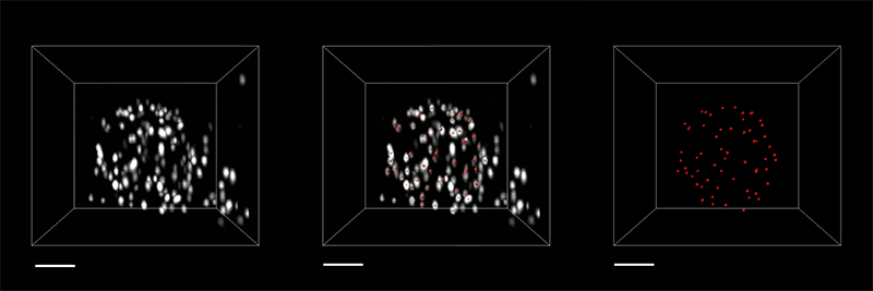
“We can search at how the preparations of these proteins alter as they are expanded and appraise how close they are to the spherical condition. Which is how we validated it and decided how faithfully we can protect the nanostructure of the styles and the relative spatial preparations of these molecules,” Ruixuan Gao claims.
Solitary molecules
The researchers also used their new hydrogel to grow cells, which include human kidney cells and mouse mind cells. They are now operating on approaches to improve the accuracy to the place wherever they can picture unique molecules inside such cells. One limitation on this diploma of accuracy is the size of the antibodies employed to label molecules in the mobile, which are about 10 to 20 nanometers extensive. To graphic personal molecules, the researchers would very likely have to have to build scaled-down labels or to incorporate the labels immediately after growth was entire.
They are also checking out no matter whether other sorts of polymers, or modified versions of the tetra-gel polymer, could enable them realize bigger accuracy.
If they can attain accuracy down to solitary molecules, several new frontiers could be explored, Boyden says. For illustration, experts could glimpse how diverse molecules interact with each individual other, which could shed gentle on cell signaling pathways, immune reaction activation, synaptic conversation, drug-concentrate on interactions, and numerous other organic phenomena.
“We’d adore to look at regions of a mobile, like the synapse in between two neurons, or other molecules associated in cell-mobile signaling, and to determine out how all the areas discuss to every single other,” he says. “How do they perform alongside one another and how do they go mistaken in diseases?”
The analysis was funded by Lisa Yang, John Doerr, Open Philanthropy, the Countrywide Institutes of Health, the Howard Hughes Professional medical Institute Simons College Students System, the Intelligence Sophisticated Analysis Assignments Exercise, the U.S. Military Analysis Laboratory, the US-Israel Binational Science Basis, the Nationwide Science Basis, the Pals of the McGovern Fellowship, and the Fellows plan of the Graphic and Info Investigation Core at Harvard Health-related School.