
To develop large-resolution, 3D photos of tissues this kind of as the mind, researchers often use two-photon microscopy, which requires aiming a superior-intensity laser at the specimen to induce fluorescence excitation. Having said that, scanning deep inside of the mind can be difficult due to the fact light-weight scatters off of tissues as it goes deeper, creating visuals blurry.
Two-photon imaging is also time-consuming, as it ordinarily calls for scanning personal pixels 1 at a time. A workforce of MIT and Harvard College scientists has now produced a modified model of two-photon imaging that can graphic further within just tissue and conduct the imaging significantly additional speedily than what was beforehand achievable.
This form of imaging could let researchers to extra speedily attain substantial-resolution visuals of constructions this sort of as blood vessels and particular person neurons in just the mind, the researchers say.
“By modifying the laser beam coming into the tissue, we confirmed that we can go deeper and we can do finer imaging than the former methods,” says Murat Yildirim, an MIT analysis scientist and 1 of the authors of the new review.
MIT graduate scholar Cheng Zheng and former postdoc Jong Kang Park are the direct authors of the paper, which appears nowadays in Science Improvements. Dushan N. Wadduwage, a former MIT postdoc who is now a John Harvard Distinguished Science Fellow in Imaging at the Center for Advanced Imaging at Harvard University, is the paper’s senior writer. Other authors incorporate Josiah Boivin, an MIT postdoc Yi Xue, a previous MIT graduate college student Mriganka Sur, the Newton Professor of Neuroscience at MIT and Peter So, an MIT professor of mechanical engineering and of biological engineering.
Deep imaging
Two-photon microscopy is effective by shining an intensive beam of in close proximity to-infrared mild on to a one stage inside of a sample, inducing simultaneous absorption of two photons at the focal point, exactly where the depth is the greatest. This lengthy-wavelength, reduced-energy light-weight can penetrate further into tissue with out detrimental it, enabling for imaging down below the surface area.
Nevertheless, two-photon excitation generates visuals by fluorescence, and the fluorescent sign is in the seen spectral area. When imaging further into tissue samples, the fluorescent mild scatters extra and the picture results in being blurry. Imaging a lot of levels of tissue is also really time-consuming. Using extensive-area imaging, in which an total aircraft of tissue is illuminated at when, can pace up the system, but the resolution of this strategy is not as good as that of place-by-place scanning.
The MIT team required to build a strategy that would allow for them to impression a substantial tissue sample all at at the time, though still protecting the higher resolution of point-by-point scanning. To accomplish that, they came up with a way to manipulate the light that they glow onto the sample. They use a variety of wide-industry microscopy, shining a aircraft of light-weight onto the tissue, but modify the amplitude of the gentle so that they can switch every single pixel on or off at different moments. Some pixels are lit up whilst nearby pixels remain darkish, and this predesigned pattern can be detected in the light scattered by the tissue.
“We can transform each pixel on or off by this form of modulation,” Zheng states. “If we convert off some of the spots, that makes room about every single pixel, so now we can know what is happening in each and every of the unique places.”
After the researchers get the uncooked images, they reconstruct just about every pixel employing a personal computer algorithm that they developed.
“We management the form of the gentle and we get the reaction from the tissue. From these responses, we try out to solve what type of scattering the tissue has. As we do the reconstructions from our uncooked photos, we can get a whole lot of information and facts that you are not able to see in the raw visuals,” Yildirim states.
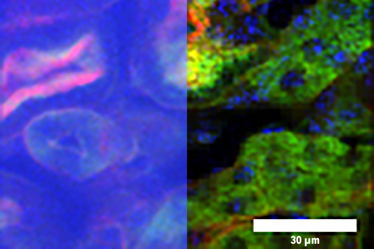
Employing this method, the researchers showed that they could graphic about 200 microns deep into slices of muscle mass and kidney tissue, and about 300 microns into the brains of mice. That is about twice as deep as was feasible with no this patterned excitation and computational reconstruction, Yildirim claims. The method can also make photos about 100 to 1,000 occasions a lot quicker than standard two-photon microscopy.
Brain construction
This type of imaging really should allow for researchers to a lot more fast acquire large-resolution illustrations or photos of neurons in the mind, as well as other structures this sort of as blood vessels. Imaging blood vessels in the brains of mice could be significantly useful for understanding a lot more about how blood flow is influenced by neurodegenerative health conditions these types of as Alzheimer’s, Yildirim states.
“All the scientific studies of blood flow or morphology of the blood vessel constructions are based on two-photon or a few-photon point scanning systems, so they’re gradual,” he says. “By using this technological know-how, we can genuinely complete higher-pace volumetric imaging of blood movement and blood vessel composition in buy to have an understanding of the variations in blood circulation.”
The approach could also lend alone to measuring neuronal exercise, by introducing voltage-sensitive fluorescent dyes or fluorescent calcium probes that light-weight up when neurons are excited. It could also be useful for examining other styles of tissue, like tumors, exactly where it could be applied to assist identify the edges of a tumor.
The research was funded by the Nationwide Institutes of Health and fitness, including the National Institute of Biomedical Imaging and Bioengineering P41 plan and the NIBIB Pathway to Independence Award, the Hamamatsu Corporation, the Samsung State-of-the-art Institute of Technological know-how, the Singapore-MIT Alliance for Investigation and Technological innovation (Intelligent), the Centre for State-of-the-art Imaging at Harvard University, and the John Harvard Distinguished Science Fellowship Method.