
In several ways, our mind and our digestive tract are deeply connected. Experience anxious may well guide to actual physical agony in the belly, even though hunger indicators from the intestine make us experience irritable. Recent scientific studies have even proposed that the micro organism living in our gut can impact some neurological ailments.
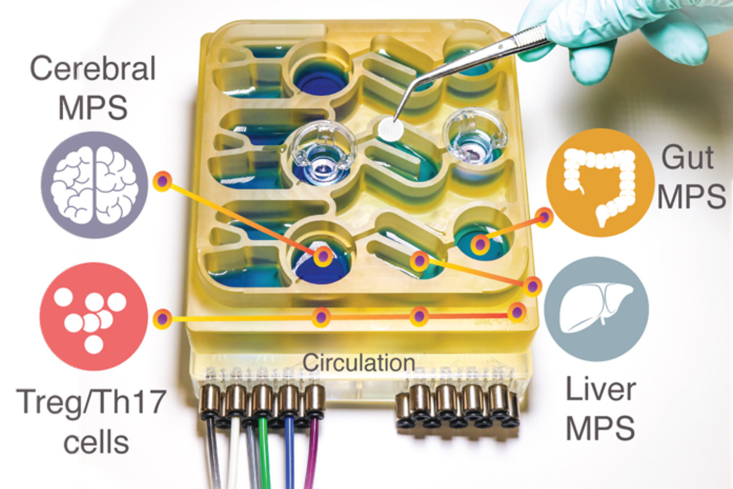
Modeling these advanced interactions in animals this kind of as mice is tough to do, simply because their physiology is pretty different from humans’. To help researchers far better recognize the intestine-mind axis, MIT scientists have made an “organs-on-a-chip” method that replicates interactions amongst the brain, liver, and colon.
Making use of that technique, the scientists were being equipped to product the influence that microbes dwelling in the intestine have on both balanced mind tissue and tissue samples derived from patients with Parkinson’s condition. They observed that limited-chain fatty acids, which are generated by microbes in the intestine and are transported to the brain, can have incredibly various results on healthy and diseased brain cells.
“While short-chain fatty acids are mostly effective to human health, we observed that underneath sure disorders they can further more exacerbate sure brain pathologies, these types of as protein misfolding and neuronal demise, connected to Parkinson’s disease,” says Martin Trapecar, an MIT postdoc and the lead author of the study.
Linda Griffith, the University of Engineering Professor of Instructing Innovation and a professor of organic engineering and mechanical engineering, and Rudolf Jaenisch, an MIT professor of biology and a member of MIT’s Whitehead Institute for Health-related Investigation, are the senior authors of the paper, which seems today in Science Innovations.
The intestine-brain link
For quite a few many years, Griffith’s lab has been producing microphysiological programs — little units that can be applied to grow engineered tissue versions of various organs, related by microfluidic channels. In some scenarios, these styles can supply much more accurate information and facts on human ailment than animal products can, Griffith claims.
In a paper released last calendar year, Griffith and Trapecar applied a microphysiological system to model interactions between the liver and the colon. In that review, they uncovered that short-chain fatty acids (SCFAs), molecules produced by microbes in the gut, can worsen autoimmune swelling connected with ulcerative colitis beneath selected situations. SCFAs, which involve butyrate, propionate, and acetate, can also have advantageous consequences on tissues, including improved immune tolerance, and they account for about 10 p.c of the energy that we get from foods.
In the new research, the MIT workforce decided to add the brain and circulating immune cells to their multiorgan method. The brain has a lot of interactions with the digestive tract, which can come about through the enteric anxious program or via the circulation of immune cells, vitamins, and hormones concerning organs.
Various years in the past, Sarkis Mazmanian, a professor of microbiology at Caltech, discovered a relationship among SCFAs and Parkinson’s illness in mice. He showed that SCFAs, which are created by bacteria as they take in undigested fiber in the intestine, sped up the development of the disease, when mice lifted in a germ-free of charge environment ended up slower to develop the ailment.
Griffith and Trapecar decided to even further investigate Mazmanian’s findings, utilizing their microphysiological model. To do that, they teamed up with Jaenisch’s lab at the Whitehead Institute. Jaenisch had previously designed a way to remodel fibroblast cells from Parkinson’s patients into pluripotent stem cells, which can then be induced to differentiate into distinct styles of brain cells — neurons, astrocytes, and microglia.
Additional than 80 p.c of Parkinson’s circumstances can not be joined to a particular gene mutation, but the rest do have a genetic lead to. The cells that the MIT researchers utilised for their Parkinson’s design carry a mutation that triggers accumulation of a protein known as alpha synuclein, which damages neurons and will cause irritation in mind cells. Jaenisch’s lab has also created mind cells that have this mutation corrected but are usually genetically identical and from the very same affected individual as the diseased cells.
Griffith and Trapecar first analyzed these two sets of mind cells in microphysiological techniques that were being not linked to any other tissues, and located that the Parkinson’s cells showed much more swelling than the wholesome, corrected cells. The Parkinson’s cells also experienced impairments in their capability to metabolize lipids and cholesterol.
Opposite outcomes
The researchers then related the brain cells to tissue versions of the colon and liver, using channels that allow immune cells and nutrients, like SCFAs, to flow in between them. They uncovered that for healthful brain cells, being exposed to SCFAs is effective, and aids them to experienced. Having said that, when brain cells derived from Parkinson’s individuals ended up uncovered to SCFAs, the useful results disappeared. Alternatively, the cells professional better stages of protein misfolding and cell loss of life.
These effects ended up viewed even when immune cells had been taken off from the process, main the scientists to hypothesize that the results are mediated by changes to lipid metabolic process.
“It appears to be that limited-chain fatty acids can be linked to neurodegenerative conditions by impacting lipid rate of metabolism rather than instantly impacting a specific immune cell populace,” Trapecar suggests. “Now the objective for us is to try out to fully grasp this.”
The scientists also plan to model other styles of neurological diseases that could be motivated by the intestine microbiome. The findings supply support for the concept that human tissue products could generate information that animal types simply cannot, Griffith says. She is now functioning on a new edition of the model that will consist of micro blood vessels connecting unique tissue styles, letting researchers to research how blood move in between tissues influences them.
“We must be really pushing development of these, simply because it is vital to start bringing much more human functions into our styles,” Griffith says. “We have been ready to get started obtaining insights into the human issue that are really hard to get from mice.”
The study was funded by DARPA, the Nationwide Institutes of Health and fitness, the Countrywide Institute of Biomedical Imaging and Bioengineering, the Countrywide Institute of Environmental Overall health Sciences, the Koch Institute Aid (main) Grant from the Nationwide Cancer Institute, and the Military Investigate Business office Institute for Collaborative Biotechnologies.